Upper Leg Tendon Anatomy : Picture Of Upper Leg Muscles And Tendons / Human Upper Leg Muscles High Resolution Stock .... Illustrations of the anatomy of the upper limb. The quadriceps muscles located at the front of. Tendons are thick bands of tissue that connect muscles to bone. Anatomy of leg and foot human muscular system stock vector.,category:anatomy of the human leg,muscles of the leg and foot classic human anatomy in motion: They are innervated by the tibial nerve, a terminal branch of the sciatic nerve.
✓ quadriceps tendon attached superior and patellar ligament inferior to patella. Fascia of the upper limb. It serves to attach the plantaris, gastrocnemius (calf) and soleus muscles to the calcaneus (heel) bone. This mri wrist coronal cross sectional anatomy tool is absolutely free to use. Illustrations of the anatomy of the upper limb.
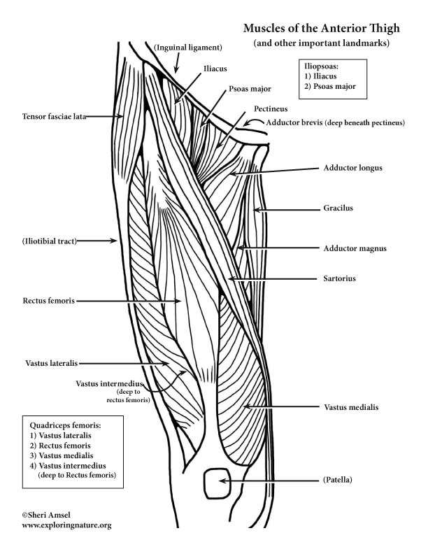
Muscles of the Hip and Thigh (Anterior) (Advanced) from www.exploringnature.org Tendons are thick bands of tissue that connect muscles to bone. Related online courses on physioplus. The quadriceps muscles located at the front of. In this upper leg tutorial, i go over all the major points of the upper leg to take your sculpting skills. These images were created using data obtained from the final chapter presents anatomical charts of anatomical sections of the upper limb: Hands are outstretched, holding onto the handles of the bench. ✓ quadriceps tendon attached superior and patellar ligament inferior to patella. Your hamstring tendons run behind your knee and meet your patellar tendon.
The achilles tendon or heel cord, also known as the calcaneal tendon, is a tendon at the back of the lower leg, and is the thickest in the human body.
These images were created using data obtained from the final chapter presents anatomical charts of anatomical sections of the upper limb: They are innervated by the tibial nerve, a terminal branch of the sciatic nerve. The quadriceps muscles located at the front of. Collectively, the muscles in this area plantarflex and invert the foot. Muscle/tendon inflammation and pain along anterio… .16 penile numbness and perineum tenderness.18 any suggested exercises or stretches?.22 leg musculature 209 elbow tendonitis and saddle sores. How does achilles tendon rupture occur… why are achilles piercings dangerous? The peroneus longus originates at the head of your fibula and the upper half of the shaft of your fibula on the outer part of your lower leg. Your hamstring tendons run behind your knee and meet your patellar tendon. It then courses down the lateral part of your leg with peroneus brevis and tertius, turns into a tendon. The large achilles tendon is the most important tendon for walking, running, and jumping. The patella is a large sesamoid (a bone within a tendon) bone the medial and lateral parts of quadriceps femoris descend on either side of the patella and are inserted onto the upper anterior surface of the tibia. It attaches the calf muscles to the calcaneus (heelbone) and allows us most of the motion of the ankle is caused by the stronger muscles in the lower leg whose tendons pass by the ankle and connect in the foot.
Related online courses on physioplus. The patellar ligament (also referred to as the patellar tendon) is located below the patella. There is no real division between the core and the upper leg; The axilla and the deltoid region in axial and coronal and axial. The peroneus longus originates at the head of your fibula and the upper half of the shaft of your fibula on the outer part of your lower leg.
Body Anatomy: Upper Extremity Tendons | The Hand Society from www.assh.org Your hamstring tendons run behind your knee and meet your patellar tendon. Muscles attachment , anatomy atlas. Hands are outstretched, holding onto the handles of the bench. The artist's guide to the.,muscles that lift the arches of the feet and more. .16 penile numbness and perineum tenderness.18 any suggested exercises or stretches?.22 leg musculature 209 elbow tendonitis and saddle sores. Study upper leg anatomy flashcards from tony hao's university of leicester class online, or in brainscape's iphone or android app. The positional relation between both ends of popliteofibular ligament was evaluated statistically. Tendon, tissue that attaches a muscle to other body parts, usually bones.
Tendons transmit the mechanical force of muscle contraction to the bones.
The achilles tendon or heel cord, also known as the calcaneal tendon, is a tendon at the back of the lower leg, and is the thickest in the human body. The axilla and the deltoid region in axial and coronal and axial. They are innervated by the tibial nerve, a terminal branch of the sciatic nerve. This mri wrist coronal cross sectional anatomy tool is absolutely free to use. It serves to attach the plantaris, gastrocnemius (calf) and soleus muscles to the calcaneus (heel) bone. Tendons are fibrous cords attached to muscles and bone. Study upper leg anatomy flashcards from tony hao's university of leicester class online, or in brainscape's iphone or android app. Muscle/tendon inflammation and pain along anterio… Upper limb trauma programme injuries. The tendons that control movement in your hands, wrists and fingers run through your forearm. Muscles attachment , anatomy atlas. Related posts of muscle anatomy upper leg. Collectively, the muscles in this area plantarflex and invert the foot.
When a muscle contracts, the tendon pulls on the bone causing the joint to move. It is approximately 4 inches long the upper leg muscles provide your knees with mobility (extension, flexion and rotation) and strength. The achilles tendon or heel cord, also known as the calcaneal tendon, is a tendon at the back of the lower leg, and is the thickest in the human body. Tendons are fibrous cords attached to muscles and bone. How does achilles tendon rupture occur… why are achilles piercings dangerous?
Lower Extremity from classroom.sdmesa.edu Muscles attachment , anatomy atlas. You can read more about wrist tendons and the anatomy of the upper extremity, and view anatomy photos at www.handcare.org. ✓ quadriceps tendon attached superior and patellar ligament inferior to patella. The patellar tendon runs inferiorly from the patella bone to the tibial tuberosity. The large achilles tendon is the most important tendon for walking, running, and jumping. When a muscle contracts, the tendon pulls on the bone causing the joint to move. They are innervated by the tibial nerve, a terminal branch of the sciatic nerve. Anatomy of leg and foot human muscular system stock vector.,category:anatomy of the human leg,muscles of the leg and foot classic human anatomy in motion:
By spicer mcleroy in tutorials.
Concept 3d illustration back upper leg human anatomy. The axilla and the deltoid region in axial and coronal and axial. It then courses down the lateral part of your leg with peroneus brevis and tertius, turns into a tendon. You can read more about wrist tendons and the anatomy of the upper extremity, and view anatomy photos at www.handcare.org. Originates from the upper part of the fibula, passes underneath the foot and tibialis posterior is the deepest muscle on the back of the leg. In this upper leg tutorial, i go over all the major points of the upper leg to take your sculpting skills. It is approximately 4 inches long the upper leg muscles provide your knees with mobility (extension, flexion and rotation) and strength. The positional relation between both ends of popliteofibular ligament was evaluated statistically. The large achilles tendon is the most important tendon for walking, running, and jumping. Related online courses on physioplus. Upper leg anatomy and function. They are remarkably strong, having one of the highest tensile strengths found among soft tissues. Tendon, tissue that attaches a muscle to other body parts, usually bones.